The lab
Research focus
- Design lab-on-chip devices for personalized global health.
- Use charge-based biomarker sensing and wireless transmission, unlike current technology.
- Support both implantable and wearable real-time diagnosis.
My work
- Ran the full fabrication process flow in the cleanroom to build GFET biosensors, from substrate to packaging to testing.
- Functionalized GFET biosensors with DNA aptamers for hormone detection.
- Co-author of a manuscript under review: Highly Sensitive and Specific Picomolar Hormone Detection in Artificial Sweat Using Aptamer-Functionalized Graphene FETs.
Project: GFET hormone biosensor
Label-free detection of estradiol and cortisol in sweat
- This project investigates a liquid-gated graphene field-effect transistor (GFET) biosensor that detects the steroid hormones estradiol and cortisol at very low (picomolar) concentrations.
- The platform combines graphene's electrical sensitivity with target-specific DNA aptamers to turn molecular binding events into measurable changes in transistor behavior.
- The long-term goal is a compact, reagent-free platform for noninvasive hormone monitoring from sweat, reducing reliance on conventional lab-based analysis.
- The system achieves picomolar-scale detection in both PBS and artificial sweat.
Motivation
- Hormones such as cortisol and estradiol carry important information about physiological and endocrine activity, but conventional measurements rely on blood samples and centralized lab techniques.
- Sweat is an attractive alternative because it can be collected noninvasively.
- Biomarker concentrations in sweat can be much lower than in blood or urine, so detection has to be highly sensitive.
- The project has three goals:
- Detect steroid hormones at picomolar concentrations.
- Keep molecular selectivity in biologically relevant solutions.
- Develop a sensing architecture that could eventually go into a portable point-of-sample system.
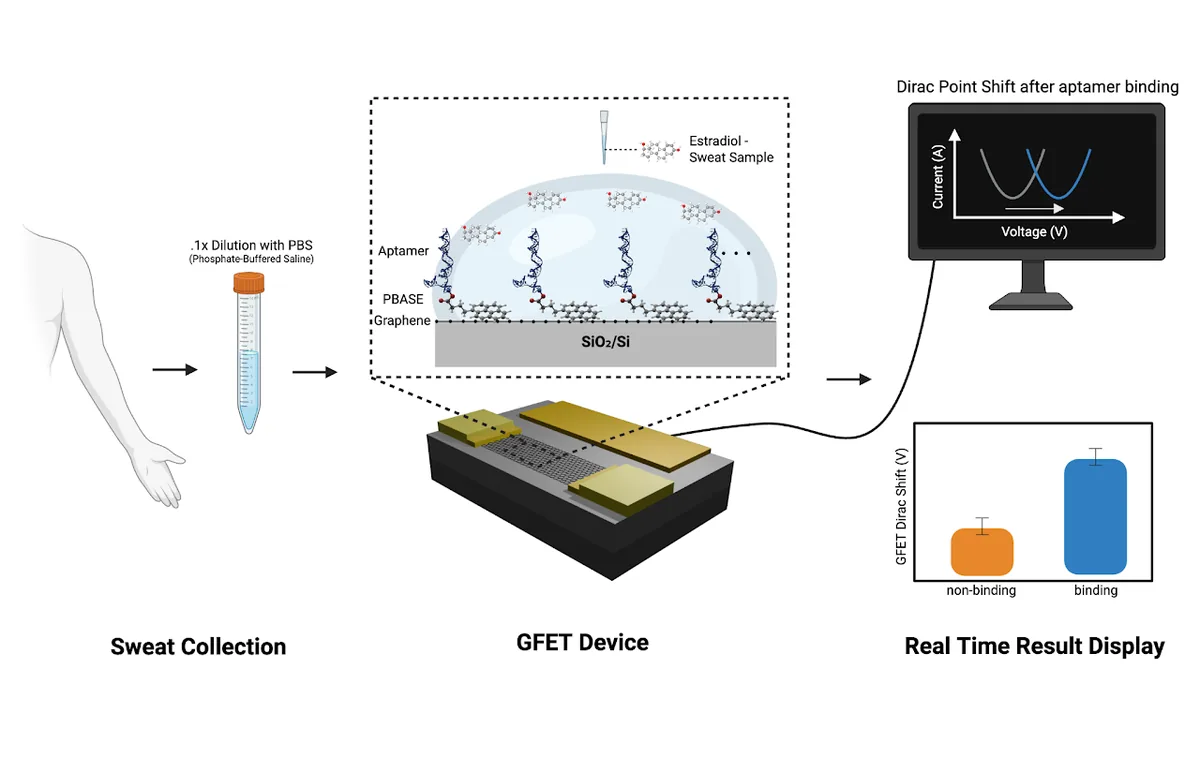
How the sensor works
The GFET is a liquid-gated transistor whose graphene channel is directly exposed to the sensing solution. The device physics page covers this in detail.
Sensing mechanism
- Hormone is introduced
- Hormone binds to the aptamer
- Aptamer conformation and local charge environment change
- Electrostatic environment near the graphene changes
- Graphene carrier concentration changes
- Dirac point shifts
A larger hormone-dependent shift indicates a stronger electrical response. A shift of about 50–100 mV is considered significant.
Biofunctionalization
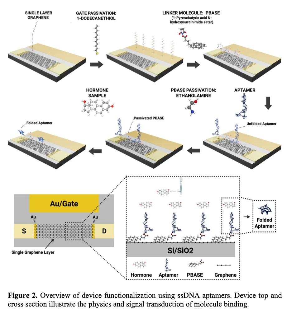
After fabrication, the GFET is chemically modified so the graphene surface can selectively recognize the target hormone.
1 · Gold gate passivation (DDT)
- The exposed Au gate is coated with a dodecanethiol (DDT) self-assembled monolayer, which forms an Au–S interface.
- DDT reduces nonspecific adsorption, parasitic electrochemical effects, and baseline drift during liquid gating.
- It adds electrical stability while preserving GFET transconductance.
2 · Graphene functionalization (PBASE)
- The graphene is modified with 1-pyrenebutyric acid N-hydroxysuccinimide ester (PBASE), the linker between graphene and aptamer.
- Pyrene group → graphene: the pyrene end attaches through π–π stacking, so functionalization doesn't disrupt graphene's sp² carbon lattice.
- NHS ester → aptamer: the NHS ester reacts with the amine-modified aptamer, making a stable attachment to the GFET surface.
3 · DNA aptamer functionalization
- Target-specific 5′-amine-modified ssDNA aptamers are immobilized on the PBASE-functionalized graphene. The aptamer is the molecular recognition element.
- When the target hormone binds, the aptamer rearranges at the nanoscale near the graphene interface.
- The resulting change in local electrostatic potential modulates the channel and shifts the measured Dirac voltage.
4 · Ethanolamine blocking
- Passivates active PBASE molecules that didn't bind an aptamer.
- Prevents unwanted binding of target hormones directly to PBASE.
Experimental testing
Two test environments
- 0.1× PBS (phosphate-buffered saline), diluted with ultrapure water to reduce Debye screening. This controlled electrolyte is used to characterize baseline stability, dose response, detection limit, selectivity, hysteresis, and device-to-device variation.
- 0.1× artificial sweat, a more biologically relevant solution with the ionic and molecular components found in sweat. It shows whether sensing still works outside a simple buffer.
Measurement procedure
- Baseline stabilization
- Transfer curve
- Hormone exposure
- Incubation
- Rinse
- Transfer curve
- Dirac voltage shift
- Baseline stabilization. Baseline solution goes into the GFET well and the device stabilizes to reduce drift in the initial Dirac voltage. Several baseline transfer curves confirm the response is stable and repeatable.
- Hormone exposure. Hormone solutions are added in increasing concentrations from the picomolar to nanomolar range, with an incubation period after each addition for the hormone–aptamer interaction.
- Rinsing. The GFET is rinsed with baseline solution to remove unbound molecules, so the signal reflects hormones bound to the aptamer surface.
- Electrical measurement. The Ids–Vgs transfer curve is measured in baseline solution after each exposure. Steps 2–4 repeat for each increasing concentration.
Signal analysis
Individual GFETs start with different electrical characteristics, so each device is evaluated against its own baseline:
ΔVDirac = VDirac(hormone, post-rinse) − VDirac(baseline, averaged)
Selectivity testing
The estradiol sensor's response is compared across estradiol, progesterone, and cortisol. The cortisol sensor is tested the same way.
Example data: estradiol and cortisol detection
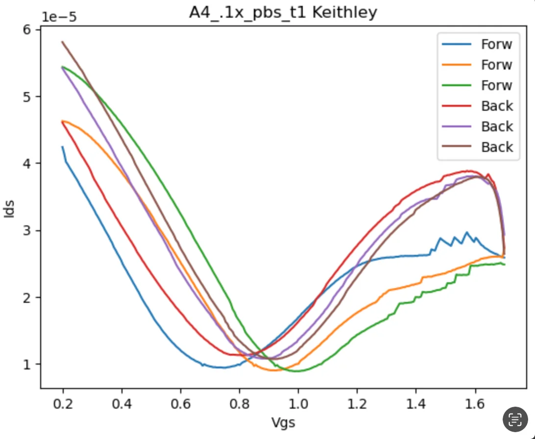
Transfer curves and Dirac voltage
- I measured source–drain current (Ids) on a probe station while sweeping gate voltage (Vgs) in forward and backward directions.
- The current minimum marks the Dirac point, graphene's characteristic charge-neutrality point.
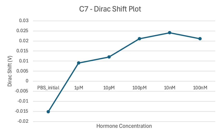
Dirac voltage shift vs. hormone concentration
- The Dirac voltage shift is plotted relative to the PBS baseline. The shift reflects hormone binding to the aptamer-functionalized channel.
- The response rises from 1 pM and saturates above about 100 pM.
- This is a single-device example. Repeatability is assessed by averaging across multiple devices.
Key results
Picomolar sensitivityEstradiol and cortisol detected at low-picomolar concentrations.
Works in artificial sweatSensing held up in a more biologically relevant sweat environment.
Hormone selectivityEach aptamer responded more strongly to its target than to the steroid interferents tested.
Label-free readoutDetection uses changes in GFET electrical characteristics, not fluorescent or enzymatic labels.
The architecture combines aptamer recognition, graphene transduction, and liquid gating in a compact single-channel sensor.